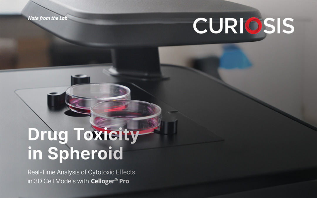
Curiosis researchers captured spheroid cytotoxic responses using CURIOSIS Celloger® Pro time-lapse imaging. Fluorescence imaging of HEK293-GFP spheroids stained with EthD-1 revealed dose-dependent shrinkage and a progressive increase in red fluorescence over time. These findings highlight the value of time-lapse imaging in enhancing drug evaluation within 3D spheroid models.
Read the full application note here
To learn more about how the CURIOSIS Celloger® Pro can enhance your automated live cell imaging workflows contact Capella Science today on (02) 9575 7512 or sales@capellascience.com.au
For research use only and not for use in diagnostic procedures.
Recent Comments